ABSTRACT
Nasopharyngeal angiofibroma is a highly vascular tumor seen typically in young adolescent males. It is an uncommon tumor accounting for less than 0.5% of all head and neck tumors. This peculiar tumor is thought to arise from the area of the sphenopalatine foramen and it classically presents with progressive nasal obstruction and epistaxis. This is a retrospective study of angiofibroma treated at our hospital over 3 years. The study discusses about clinical features, management options and the role of embolization in reducing the intra-operative blood loss.
KEYWORDS
Angiofibroma; Embolization; Nasopharynx; Vascular
Introduction
Juvenile Nasopharyngeal Angiofibroma is a rare tumor occurring almost exclusively in young males. Though the tumor is considered benign, it has a propensity for local invasion and destruction. It is classically described as invading “through natural fissures and foramina”. The pathogenesis of the angiofibroma has been a topic of much debate, but most authors agree that the tumor arises from the posterolateral wall of the nasal cavity and the adjoining supero-lateral nasopharyngeal wall and the sphenopalatine foramen is invariably involved. The tumor characteristically presents as progressive unilateral nasal obstruction and epistaxis. Diagnosis is made by clinical examination and imaging studies. Surgery is the gold standard treatment for angiofibroma [1]. Our study focuses on the presentation, surgical approach and more importantly the role of preoperative embolization in managing these highly vascular tumors.
Materials and Methods
The following study is a retrospective study done at ENT department in Government ENT Hospital, Hyderabad, India. From May 2010 to June 2013, 30 patients of angiofibroma were operated, and are included in this study.
Discussion
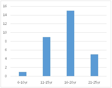
All the 30 patients were males and no female case is reported in our series. The study includes patients between age of 9 and 24 years with a mean age of 16.2 years [Chart 1]. 15 patients fell in the most common age group of 16-20 years. Among all the symptoms epistaxis and nasal obstruction were the predominant symptoms in our study. Epistaxis was the most troublesome symptom for which the patients took medical advice. Amount of bleeding varied from 20-150 ml in each episode, which was spontaneous. 2 patients required post nasal packing in the emergency room to stop the bleeding. Nasal obstruction was progressive and much better tolerated symptom than epistaxis as majority of our patients presented with unilateral obstruction initially (Table 1). Nasal discharge was seen in 11 patients. 2 patients both recurrent cases, presented to us with isolated cheek swelling.
The tumor was evaluated by clinical examination, diagnostic nasal endoscopy and imaging. CT scan of nose and paranasal sinuses was performed in all the cases. The extension of the tumor was studied. In our study we applied the Fisch staging system of the angiofibroma [2] (Table 2).
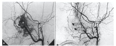
The goal of tumor embolization is to occlude selectively the External Carotid Artery (ECA) feeders through intratumoral deposition of embolic material [3]. The embolic agents in common use are Polyvinyl Alcohol (PVA), Embospheres, liquid embolic agents (glue, Ethyl Vinyl Alcohol Copolymer [EVOH], or Onyx [ev3, Irvine, Calif]), gelatin sponge (Gelfoam; Phadia), and coils. The embolization is ideally performed 24 - 72 hours before surgical resection to allow maximal thrombosis of the occluded vessels and prevent recanalization of the occluded arteries or formation of collateral arterial channels (Figure 1).
The most common complication of embolization is facial pain. Serious complications occur in less than two percent of patients. These are usually related to particle reflux, poor technique, or non-visualization of dangerous anastomoses resulting in blindness or irreversible neurologic deficits [4].
Results
Nasopharynx was the most common site of extension in our study, with 22 patients having a nasopharyngeal extent of the tumor. Fifteen patients had Fisch II tumor at presentation. Two tumors had to be upstaged intraoperatively (case nos. 6 and 26).
Among the 30 cases, pre-operative embolization with gelatin sponge (Gelfoam) was performed in 11 cases in our study. Embolization was performed by interventional radiologist 48 hrs prior to surgery in all the eleven patients. All the patients tolerated the procedure well, with 2 patients having facial pain for 2 days. Internal maxillary artery was the main feeding vessel in 9 patients and in 2 patients both internal maxillary and ascending pharyngeal artery.
Based on the clinical and imaging findings, surgery was planned for the all patients. Different approaches were used for the surgery depending on the extent of the tumor. Four main approaches used in this study are transpalatal, endoscopic, lateral rhinotomy and combined approach (Transpalatal and Endoscopic) [5]. Weber Ferguson approach was done in 2 patients.
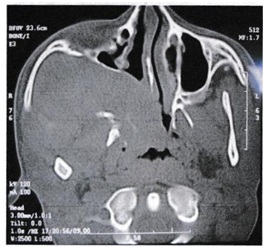
Among the eleven embolized patients, the intraoperative blood loss in 4 patients was insignificant and in the rest of patients ranged from 250-700 ml with an average of 450 ml [3-5]. Embolization reduced intraoperative blood loss from an average of 950 ml in the non-embolized patients to 380 ml in the embolized patients. Lateral rhinotomy approach was performed in 6 patients, and this approach gives excellent exposure in infratemporal fossa involvement (Figure 2). Weber Ferguson approach was done in 2 patients with stage IIIa and IIIb.
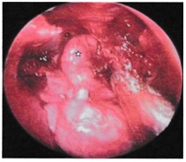
Endoscopic trans-nasal approach was performed in eleven patients [6,7]. This approach gives excellent exposure in stage I and stage II tumor (Figure 3).
All thirty patients were followed up for a period of two years (Table 3). Diagnostic nasal endoscopy was performed each month and a CT scans after one year. Recurrence was seen in three patients (case no. 3, 17 and 29) in the series. One patient who had been embolized and the other two, embolization was not performed. These patients presented with epistaxis after a mean duration of 4 months. All the three patients were later operated by endoscopic approach.
Conclusion
Embolization pre-operatively reduces the blood loss, need for blood transfusions and decreases morbidity. Endoscopic approach obviates the need for an incision and gives excellent exposure in selected cases.
Figures
Chart 1: 06-10 yrs [1]
11-15 yrs [9]
16-20 yrs [15]
21-25 yrs [5]
Summary of the age group of nasopharyngeal angiofibroma. Numbers in bracket indicate number of patients.
Figure 1: (a) Before embolization (tumor blush can be seen) (b) After embolization.
Figure 2: CT Scan showing extension of angiofibroma into the infratemporal fossa.
Figure 3: Endoscopic transnasal approach in a stage II tumor (embolized), intra-operative blood loss was insignificant.
Tables
| Symptoms/Signs | No. of Patients | Percentage (%) |
| (n=30) |
| Nasal obstruction | 22 | 73 |
| Epistaxis | 19 | 63 |
| Nasal discharge | 11 | 36 |
| Change in voice | 8 | 26 |
| Headache | 6 | 20 |
| Snoring | 5 | 16 |
| Face deformity | 3 | 10 |
| Cheek swelling | 2 | 6 |
| Intra-oral swelling | 2 | 6 |
Table 1: Clinical presentation of angiofibroma.
| Stage I | Tumor limited to nasopharyngealcavity, bone destruction negligible or limited to sphenopalatine foramen |
| Stage II | Tumor invading the pterygopalatine fossa or the maxillary, ethmoid or sphenoid sinus with bone destruction |
| Stage III | Tumor invading theinfratemporal fossa or orbital regiona) Without intracranial involvementb) With intracranial extradural involvement (parasellar) |
| Stage IV | Intracranial intradural involvementa) Without infiltration of the cavernous sinus, pituitary fossa or optic chiasmb) With infiltration of the cavernous sinus, pituitary fossa or optic chiasm |
Table 2: Fisch staging of juvenile nasopharyngeal angiofibroma.
| Case No | Age | Pre-op stage | Embolization | Surgical approach |
| 1 | 24 | IIIa | No | Lateral Rhinotomy |
| 2 | 17 | II | No | Transpalatal |
| 3 | 16 | II | No | Endoscopic Transnasal |
| 4 | 21 | IIIb | No | Lateral Rhinotomy |
| 5 | 15 | II | No | Endoscopic Transnasal |
| 6 | 13 | II * | No | Lateral Rhinotomy |
| 7 | 16 | II | No | Endoscopic/Transpalatal |
| 8 | 18 | II | Yes | Endoscopic Transnasal |
| 9 | 20 | IIIa | Yes | Endoscopic/Transpalatal |
| 10 | 13 | II | Yes | Endoscopic |
| 11 | 20 | IIIa | Yes | Lateral Rhinotomy |
| 12 | 17 | I | No | Transpalatal |
| 13 | 1 | I | No | Transpalatal |
| 14 | 20 | II | Yes | Endoscopic Transnasal |
| 15 | 14 | IIIb | Yes | Weber-Ferguson |
| 16 | 20 | II | No | Endoscopic Transnasal |
| 17 | 16 | II | No | Transpalatal |
| 18 | 15 | IIIa | Yes | Endoscopic/Transpalatal |
| 19 | 13 | II | No | Endoscopic Transnasal |
| 20 | 15 | IIIa | No | Lateral Rhinotomy |
| 21 | 17 | I | No | Transpalatal |
| 22 | 17 | II | Yes | Endoscopic Transnasal |
| 23 | 19 | II | No | Endoscopic Transnasal |
| 24 | 22 | I | No | Endoscopic Transnasal |
| 25 | 18 | IIIa | Yes | Lateral Rhinotomy |
| 26 | 23 | II * | No | Transpalatal |
| 27 | 15 | II | No | Endoscopic Transnasal |
| 28 | 15 | IIIa | No | Weber-Ferguson |
| 29 | 9 | II | Yes | Endoscopic-Lateral Rhinotomy |
| 30 | 23 | II | Yes | Transpalatal |
Table 3: Summary of age group, pre-op stage, embolization and surgical approach in our study.
*- Upstaged intraoperatively