Palatal Aponeurosis Massage: A Diagnostic and Treatment Tool to Tinnitus and Otological Symptoms
*Corresponding Author(s):
Claudia Barros CoelhoDepartment Of Otolaryngolgy, UNIVATES College Of Medicine, Lajeado, Brazil
Tel:+55 5137102610,
Email:claudia.coelho@univates.br
Abstract
The number of patients seeking treatment for aural fullness, increase in sound threshold, hyperacusis, and tinnitus-together diagnosed as Tonic Tympani Tensor Syndrome (TTTS) as increased greatly during the Covid 19 pandemic. These subjects often present with complaints of Temporomandibular Joint (TMJ) disorders, bruxism, and anxiety. Herein, we report a novel diagnostic and treatment method. This series of cases (n=22) was collected from consecutive patients who attended a tinnitus clinic between February and July 2021. After a clinical history suggestive of TTTS and associated TMJ disorder with myofascial pain and stomatognathic muscle tenderness, subjects were submitted to a novel myofascial relaxing and stretching technique applied to the palatal aponeurosis. Treatment was performed in a single session. The outcome measurement was based on a tinnitus loudness rating scale of 0-100. The mean tinnitus loudness score before treatment was 54.77 (confidence interval-CI 45.8 to 65.47) and after treatment 25.2 (CI 19.59-to 32.61) (p < 0.001). The magnitude effect mean ratio was 0.46 (95% CI .0.39 to 0.54.), p < 0,001.
The massage reduced tinnitus loudness immediately after being performed. It also reduced associated otological complaints such as ear pressure and muffled hearing.
A novel clinical treatment protocol composed of palatal aponeurosis massage showed promising results in subjects with tinnitus arising from a TMJ disorder with a myofascial disorder. Further research with objective measurements in a controlled clinical trial is necessary to validate this method. This could have a significant impact on the management of this subset of patients.
Keywords
Covid19; Temporomandibular joint disorder; Tensor tympani; Tensor veli palatine; Tinnitus; Tinnitus loudness; Tinnitus treatment; Tonic tensor tympani syndrome; Palatal aponeurosis; Somatosensory tinnitus
Introduction
Tinnitus is a worldwide common symptom, affecting millions and yet, for most cases, there is no available cure [1]. Moreover, treatment options that have been evaluated in clinical trials aim to mitigate the impact on a patient’s life rather than offer hope for a cure [2]. Probst et al. note that increases in tinnitus loudness result in an increase in individual stress. Thus, therapeutic interventions such as sound therapy and cognitive behaviour therapy that reduce only the general impact level, without changing the tinnitus loudness, have strong limitations.
Tinnitus has many different causes and probably there are many different mechanisms. Any change compromising the auditory system, in any location, can lead to tinnitus. The nature of tinnitus, as a highly complex condition, presents many different subtypes that should be identified [3] adding to the difficulty of finding a cure. Appreciating and understanding different subtypes will help lead the way to different treatments, and will be critical for exploring and finding cures for different subtypes of tinnitus [4].
During the COVID-19 pandemic we have observed a large new set of tinnitus patients in our clinic who complain of tinnitus, ear pain, ear pressure, muffled hearing, and myofascial pain. Some describe a change in the characteristics of their existing tinnitus while others describe the recent appearance of tinnitus that they previously did not have. Psychological issues involved during the pandemic have also increased the number of individuals presenting TMJ disorder and bruxism [5].
Somatosensory tinnitus
Some people perceive a change in tinnitus intensity associated with muscle contractions, mechanical pressure on myofascial trigger points, cutaneous stimulation, or joint movements. This is called Somatosensory Tinnitus (ST), which is a subtype of tinnitus, present in about 12% to 43% of tinnitus patients. ST mechanisms are based on neural connections and integration between the auditory and somatosensory pathways in the DCN, inferior colliculus, and auditory cortex [6-9]. Altered input affecting the trigeminal pathway such as observed on Temporomandibular Disorders (TMDs) or bruxism could change the loudness perception of an existing tinnitus or generate tinnitus.
TMJ disorders
TMJ disorders are a set of heterogeneous craniocervical pathologies that alter the functioning of the muscles of mastication, cervical muscles, and the temporomandibular joint, among other associated structures. These disorders, generally, do not have the same etiology, but are grouped in the same diagnosis, as they act in the orofacial structures in a similar way. These changes affect the entire musculature of the face, neck, and head, in addition to having a close connection with the auricular system. Malfunctioning of the muscles in the region can cause orofacial pain and cause very diverse secondary otological symptoms, such as otalgia, tinnitus, vertigo and hearing loss [10].
Bruxism
Bruxism, a repetitive jaw-muscle activity characterized by clenching or grinding of the teeth and/or by bracing or thrusting the mandible [11], plays a significant role in TMJ disorder. Stress and psycho-emotional tension can affect the function of the masticatory system, inducing several pathological reactions. If the muscle tension persists, structural dysfunctions, such as hypertrophy, tenderness, and activation of trigger points, causing myofascial pain on the stomatognathic muscle may occur [12].
Auditory system and TMJ disorders
TMJ disorder and bruxism disorder have an important effect on the auditory system [13]. The relationship between otological symptoms such as tinnitus and TMJ disorders have been related by Costen almost a century ago and republished [14]. However, the link is still not clearly established. Costen described a combination of various otological symptoms-subjective hearing loss, headache, otalgia, and otic fullness or blocked ear sensation, vertigo, and/or tinnitus-in a series of patients. These findings were described by many other authors (for a comprehensive review see Ramirez) [15].
The muscles of mastication, jaw, and middle ear bones (malleus, incus, and stapes anterior pillar) develop from the first pharyngeal arch or Meckel cartilage. They are innervated by a branch from the trigeminal nerve (CN V) motor nucleus, as well as the masseter temporalis, medial pterygoid, lateral pterygoid, mylohyoid, and the anterior belly of digastric muscles. This cluster of V motoneurons, in addition to the tympanic tensor and tensor veli palatine, activate muscles of mastication. They are also active in cranial ventroflexion and deglutition [16].
Anatomically, the Tympanic Tensor (TT) and the Tensor Veli Palatine (TVP) are muscles of the middle ear and comprise the “strange chewing” muscles capable of causing otological symptoms by TMD due to common motor innervation. These muscle complexes are part of the normal physiology of hearing but are also responsible for secondary and inexplicable otic referred symptoms when TMJ disorder is present [17].
The intertwined ontogeny, anatomy, and physiology of these muscles can lead to symptoms of TMJ disorder producing constant (spastic) or episodic (clonic) contraction. Constant tension in the TVP and TT muscles can lead to fatigue and secondary symptoms referred to the ear, especially when there is a dysfunctional stomatognathic state [15]. This dysfunction might be a result of TMJ disorder and bruxism, leading to the onset of myofascial trigger points and shortening of the TVP and TT muscular length.
The TVP and TT muscles play an important role in regulating hearing. The former is mainly responsible for the opening of the auditory tube and the latter inserts on the malleus, acting to tense the tympanic membrane (hence its name, tensor tympani) [15]. Mechanically and neurologically, the TT and the TVP work in a synergistic way. Both muscles contract simultaneously during swallowing, producing an internal deflection of the tympanic membrane and opening the Eustachian tube to ventilate the middle ear [18]. In figure 1, one can see the common tendon that connects the TVP and TT. The primary role of the TT is to protect the cochlea from noise trauma. The acoustic reflex is a neurological control loop that increases tension in the tympanic membrane and protects the inner ear from harmful sound levels [19-21]. Besides that, the tensor tympani muscle allows the hearing discrimination of low frequencies and work as a barometric pressure receiver in the middle ear Eustachian tube opening reflex [17,22].
 Figure 1: Anatomic dissection demonstrating the anatomical connection between the TT and the TVP in a human temporal bone. 1. TVP, 2. TT, 3. Stapes, 4. Stapedial muscle tendon, 5, VII CP, 6. Chorda tympani nerve, 7. Auditory tube opened, 8. Tendon and muscular connection linking TVP and TT (from Dr Ramirez Aristeguieta personal dissection collection).
Palatal aponeurosis
Palatal Aponeurosis (PA) is a flexible and fibrous membrane attached to the posterior edge of the hard palate and extends from the Tensor Veli Palatini (TVP). It plays a key role in the palatopharyngeal function in preserving the shape of the soft palate and in the transmission of muscular force to the soft palate and pharyngeal muscles [23]. In a recent study [24] it was possible to identify that the TVP extends like a tendon, bypassing the pterygoid hamulus to become the PA, leaving a space between the aponeurosis and the hamulus. With a mechanism similar to a pulley, the PA slides around the pterygoid hamulus, helping the musculature elevating the soft palate. The authors also suggest that the PA, as part of the TVP, supplies tension and controls the opening and closing of the Eustachian tube.
Tonic Tensor Tympani Syndrome (TTTS)
TTTS is characterized by a constant tonic contraction of the TT, generating symptoms such as tinnitus and hyperacusis, ear pain, fullness and dizziness. The clinical scenario is similar to Meniere's syndrome and is also, when a TMJ disorder is present, responsible for a range of secondary and inexplicable otological symptoms [25-27]. The etiology of TTTS is not yet evident, but there is a dysregulation in the reflex control of the muscle, causing continuous contraction. Clinical features include otalgia, ear fullness, tinnitus (usually at low sensation level and sometimes pulsating, dysacusis (sensations such as murmurs, clicks, tickling), tension headache, and vertigo. Patients may complain of difficulties on sound discrimination like what is seen on stapedial paralysis after head trauma [25].
The sustained contraction of TT muscle might alter the spatial arrangement of the middle ear ossicles and can lead to a rise in endolymphatic pressure through the transmitted changes from the oval window to the fluids of the labyrinth. The same middle ear pathogenic muscle mechanism can diminish the transmitting vibration from the tympanic membrane, resulting in a hearing impairment [28]. As indicated previously, Shapiro and Truex [29] suggested that this might lead to vestibular and cochlear imbalance and similar symptomatic reactions as in Meniere’s syndrome.
Development of the palatal aponeurosis massage
As tinnitus researchers, one of our objectives is to investigate possible treatments to mitigate tinnitus. Particularly, we are interested in finding a treatment that could reduce tinnitus loudness. This has led to our interest in TTTS and exploring further ways of diagnosing and treating this entity.
Based on anatomical dissections by Ramirez and Zhao et al. [15,23] which has shown an intimate anatomical and functional unity between the TT and TVP and PA, and also, on case report [30] that used osteopathic technic to treat objective tinnitus, we have develop a novel technique, using the palatine aponeurosis as a route for the TT. If a muscle has become tense, there should be a way of releasing the tension. We started to palpate and massage the palatal aponeurosis, using the pterygoid hamulus as a landmark. We have performed this technique in patients presenting symptoms of TTTS and associated tenderness of the masticatory muscles and trigger points. We saw an immediate release of tension in the area, and, remarkably, followed by a decrease in tinnitus loudness and subjective hearing improvement. Patients would say that their ears have opened, and they could hear better. In addition, we observed a remission of ear pain when this was present. We have named this new technique the Palatal Aponeurosis Massage (PAM).
The aim of this series of case study is to evaluate the effect of the PAM on tinnitus loudness on subjects with presence of temporomandibular disorders with myofascial pain. Our main outcome is the perceived loudness of tinnitus on a scale of 0-100 (0=no tinnitus, 100=loudest tinnitus you can experience [31].
Our null hypothesis is that the mean value of tinnitus loudness is the same before and after the procedure. Our alternative hypothesis is that there is a significant change on the mean value of tinnitus loudness after the procedure.
Materials And Methods
We invited patients attending the main author’s tinnitus clinic, in their first appointment, to participate on this preliminary trial if they had tinnitus associated to TMD symptoms according to The Diagnostic Criteria for Temporomandibular Disorders [32] with local myalgia.
Inclusion criteria: Presence of tinnitus, reported pain in masticatory muscles, pain on palpation on temporalis or masseter muscle in at least 3 sites, one of them at least in the same side of the reported pain.
Exclusion criteria: presence of external and middle ear infection, structural and functional palatal anomalies (palatal clefts, including submucosal clefts, velopharyngeal dysfunction), overly sensitive pharyngeal reflex and reduced mouth opening preventing to perform the intervention.
To participate in this study, all subjects gave written consent to perform the procedure, after a clear explanation that it was a novel technique, which was not a formal treatment, and we were collecting information about their experience.
Before the intervention took place, we asked the subjects to rate their tinnitus loudness as proposed by Tyler et al. [31]. “I want you to rate the loudness of your tinnitus on a scale from 0 to 100; 0 representing no perception of tinnitus and 100 representing the loudest tinnitus you can experience”. Intermediate numbers represent intermediate loudness ratings.
With the subject on a sitting position inside an audiometric booth, the examiner, wearing gloves and with short nails, introduces the 2nd finger towards the palatine aposeurosis above the pterygoid hamulus, while holding the back of the neck with the opposite hand (Figure 2). Gently, a 15-second digital pressure on the horizontal plane over the palatal aponeuroris intercalating with a digital pressure over longitudinal plane lateral to the pterygoid hamulus was applied. The pressure intensity will be determined by the subject’s tolerability, which increases over the procedure. It is possible to feel a smoothing effect over aponeurosis and the tendon, and sometimes a twitch response.
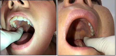 Figure 2: Palatal Aponeurosis Massage over the hamulus pterygoid: (a) on the right (b) on the left sides.
The procedure is carried out once on each side and after that it is repeated two more times, resulting in 6 digital pressures in total. The health-care professional explains to the patient the need to keep the jaw and muscles relaxed, without pressing the teeth, and that she/he might feel a slight discomfort and possibly a vomiting reflex. Some lacrimation might occur in the ipsilateral side of the massage. Local anesthetics were not used on this trial.
Once the procedure was completed, we asked the subject to rate their tinnitus loudness again.
Statistical analysis
As this is an exploratory and preliminary study, we did not perform a formal sample size calculation.
Quantitative data were described as mean, Standard Deviation (SD), median, Interquartile Range (IQR) (P25-P75) and minimum and maximum values. Categorical information was expressed by counts and percentages.
To compare the values of the Tinnitus magnitude rating scale between the baseline and after the procedure, we used a generalized Estimating Equation (GEE) model with a link function based on the negative binomial distribution with an exchangeable correlation matrix. Quasi Likelihood under Independence Model Criterion (QIC) value was 29.405. The magnitude of the effect was expressed by the ratio of the means with their respective 95% confidence interval. P values below 0.05 were considered statistically significant. Data were analyzed using the IBM-SPSS version 27.0 program.
Results
The procedure was applied on 34 consecutive subjects with tinnitus and the presence of TMJ disorder, in their first appointment at the first author’s tinnitus clinic, between February and July 2021. As the learning curve improved, and new insights were obtained, we began to collect data in a more detailed way and the first 12 subjects were excluded from the analysis. The final sample counted on 22 subjects. The procedure was performed by the first author in all subjects
The sample counted 11 females and 11 males; ages ranged from 29 to 69 years (mean 54 years median 55.5 years SD±10.5years). Tinnitus location was generally unilateral, 68.2% (n=15) similarly between the right and left ears. Most common associated symptoms included aural fullness, muffled hearing, hyperacusis, dizziness, facial and ear pain. All subjects presented masticatory muscle tenderness and pain during palpation, mostly the masseter and temporalis muscles.
Tinnitus loudness scale rating
The distribution of our primary outcome measurement is shown on figure 3. Tinnitus loudness scale before treatment had a mean value of 54.77 points, median 55, SD±23.9. For the primary outcome of the study, figure 3 presents the individual scores before and after treatment.
 Figure 3: Scatter plot represents tinnitus loudness scores before and after the Palatal Aponeurosis Massage. The additional box plots describe the Mean, Standard Deviation (SD), Median, Interquartile range (IQR) and number of subjects (n).
Changes on loudness scores were highly significant when comparing loudness scores with the GEE model with a link function based on the negative binomial distribution with an exchangeable correlation matrix based on paired two-tailed t tests.
The mean value of tinnitus loudness score before treatment was 54.77 (Confidence interval-CI 45.8 to 65.47) and after treatment 25.2 (CI 19.59-to 32.61), (p< 0.001). The magnitude effect of the intervention was measured by the mean ratio which was 0.46 (95% CI .0.39 to 0.54.), p<0,001.
We explored the effect of age, sex, and tinnitus location on the magnitude variation of treatment response. We did not find any significant findings on response regarding these factors: Sex: Mean ratio 0.466 (CI 95% 0.395-0.550); tinnitus location mean ratio 0.507 (CI 95% 0.407-0.631); age mean ratio 0.399 (CI 95% 0.193- 0.827). Changes on mean loudness scores for each of these factors are described on table 1.
|
Factor |
Meanloudness |
Confidence Interval 95% |
p-value |
|
Unilateral |
B 55 |
46-66 |
0.9 |
|
A 25 |
19-32 |
||
|
B 53 |
35-81 |
||
|
A 25 |
13-46 |
||
|
Male |
B 63 |
58-69 |
0.3 |
|
A 27 |
20-35 |
||
|
B 45 |
31-66 |
||
|
A 23 |
14-36 |
||
|
≤55 y |
B 46 |
32-66 |
0.6 |
|
A 21 |
13-36 |
||
|
B 62 |
55-71 |
||
|
A 28 |
23-35 |
Table 1: Exploratory analysis to evaluate the effect of tinnitus location, sex, and age on the mean loudness scores before (B) and after (A) the massage, y (years).
Adverse effects: Some patients experienced a gagging reflex during the procedure, which was managed by giving a rest before performing the whole set of manoeuvres. Lacrimation often occurred on the ipsilateral side of the manipulation. To minimize discomfort perceived during the procedure, we applied progressive tension on the stimulation area. All subjects tolerated the massage. Bleeding was not observed.
Discussion
The effect of the PAM on the decrease of tinnitus loudness perception has shown promising results on a subgroup of tinnitus sufferers with associated TMJD.
Here, we propose a novel approach, based on anatomical and functional unity between the PA, TVP and TT, using the PA as a route for activating the TT. The constant contraction of TVP may produce myotonic and myofibrotic adherences resulting from chronic TMD dysfunction that can be released by sustained finger manipulation on the zone described here on the PA.
We hypothesize that this manipulation can also affect the Tympanic Tensor, also releasing its tension, changing perception on otologic symptoms, including tinnitus loudness. Also, it is possible that TVP and TP relaxation could modulate the neural input of the trigeminal nerve, on DCN, inferior colliculus, and auditory cortex, and decrease a possible presentation of somatosensory tinnitus on these patients.
Another hypothesis is that PAM could be performed to help on the differential diagnosis between TTTS, superior semicircular canal dehiscence syndrome [33], benign intracranial hypertension [34,] and Meniere Disease [35] These different tinnitus etiologies might share similar symptoms such as aural fullness, autophony, hearing loss, hyperacusis and dizziness.
With a literature review we found the “Trigeminal Pharyngioplasty Procedure” (TPP), which is also based on the embryology and anatomy of TVP and TT muscles. TPP was developed at White Memorial Medical Center’s Craniofacial Pain/TMJ Clinic in 1994 and published by Schames et al., [36]. TPP was applied to release the dysfunction of these muscles myocontraction, and consequently, sustained myofibrosis (myofascial trigger points with shortening of the muscles) because of parafunctional activities of bruxism resulting in significant TMD burden to the masticatory musculature and to the TMJ complex. Patients with facial pain, and symptoms like ear pain, pressure in the ear, vertigo, and tinnitus and hearing impairment had 75% rate of success in a complete or partial improvement of their condition with the treatment. TPP, as described by the authors, consists of “sliding the index finger medially past the retromolar pad in the oral cavity to the posterior aspect of the soft palate, slip underneath the soft palate and to slide in a posterior superior lateral direction into the Fossa of Rosenmüller. Gently sliding the index finger in an inferior lateral direction along the torus tubarius at the orifice of the Eustachian tube. This finger-lysing any adhesions and massaging the muscle area”.
TPP is different to our PAM, and it has possible risks, as described by the authors, including possible damage to carotid aneurysm (if present), the need to anesthetize the oro-pharynx in some patients, resulting in difficulty swallowing. Also, the presence of a gag reflex from the discomfort of having a gloved finger inserted in the back of throat, can cause pain, irritated throat, and bleeding.
Study Limitations
There are several limitations on this preliminary study. Our sample was performed in a single centre, we had no controls, and subjects were not randomized. The changes perceived on tinnitus loudness were measured only by a rating scale. Measurements of tinnitus loudness and minimal masking levels could help us to measure psychoacoustic changes. Tinnitus duration still has to be explored on the magnitude variation of treatment response.
Also, on this preliminary trial we focused the effect of TVPRM on tinnitus loudness, future trials could also test its effect on tinnitus distress and hyperacusis, using proper measurements.
It would be appropriate in future research, to collect data as parameters such as tympanometry, that could register a possible change on TM compliance, wideband tympanometry, to evaluate a tympanic resonance. Otoscopic recording before and after the procedure could monitor possible changes on malleus position following the massage. Also, the referred change on hearing thresholds should be measured directly, such as audiometry and otoacoustic emissions. Long term follow up of the effect is necessary, as well if the instructions given to patients to perform the procedure at their homes would also have an effect. Further, associated treatments directed to the myofascial component of TMJ Dysfunction, could have a better effect on long term management of this subtype of tinnitus patients.
If these results could be replicated, that would provide an advance on tinnitus treatment, accessible to professionals dealing with tinnitus, patients and easily performed and costless.
Acknowledgement
We thank Dr Andrew Bell BSc, BA, MSc, PhD Australian National University, Eccles Institute of Neuroscience and Dr Robert Levine, MD, Department of Otolaryngology Tel Aviv Medical Center, for their valuable contribution on this manuscript elaboration.
Conflicts of Interest
The authors declare no conflict of interest.
References
- Cima RFF, Mazurek B, Haider H, Kikidis D, Lapira A (2019) A multidisciplinary european guideline for tinnitus: Diagnostics, assessment, and treatment. HNO: 67: 10-42.
- Baguley D, McFerran D, Hall D (2013) Tinnitus. The Lancet 382: 1600-1607.
- Richard T, Claudia C, Pan T, Haihong J, William N, et al. (2008) Identifying tinnitus subgroups with cluster analysis. American Journal of Audiology 17: 176-184.
- Coelho CB, Santos R, Campara KF, Tyler R (2020) Classification of tinnitus: Multiple causes with the same name. Otolaryngol Clin North Am 53: 515-529.
- Emodi-Perlman A, Eli I, Smardz J, Uziel N, Wieckiewicz G, et al. (2020) Temporomandibular disorders and bruxism outbreak as a possible factor of orofacial pain worsening during the COVID-19 pandemic-concomitant research in two countries. J Clin Med 9: 3250.
- Levine RA (1999) Somatic (craniocervical) tinnitus and the dorsal cochlear nucleus hypothesis. American Journal of Otolaryngology 20: 351-362.
- Shore S, Zhou J, Koehler S (2007) Neural mechanisms underlying somatic tinnitus. Progress in Brain Research 166: 107-123.
- Levine RA, Abel M, Cheng H (2003) CNS somatosensory-auditory interactions elicit or modulate tinnitus. Exp Brain Res 153: 643-648.
- Dehmel S, Cui YL, Shore SE (2008) Cross-modal interactions of auditory and somatic inputs in the brainstem and midbrain and their imbalance in tinnitus and deafness. Am J Audiol 17: 193-209.
- Lam DK, Lawrence HP, Tenenbaum HC (2001) Aural symptoms in temporomandibular disorder patients attending a craniofacial pain unit. Journal of Orofacial Pain 15: 146-157.
- Lobbezoo F, Ahlberg J, Glaros AG, Kato T, Koyano K, et al. (2013) Bruxism defined and graded: An international consensus. Journal of Oral Rehabilitation 40: 2-4.
- Anna S, Joanna K, Teresa S, Maria G, Aneta W (2015) The influence of emotional state on the masticatory muscles function in the group of young healthy adults. Biomed Res Int.
- Shankland WE (2001) Migraine and tension-type headache reduction through pericranial muscular suppression: A preliminary report. Cranio 19: 269-278.
- Costen JB (1997) A syndrome of ear and sinus symptoms dependent upon disturbed function of the temporomandibular joint. Ann Otol Rhinol Laryngol 106: 805-819.
- Ramirez Aristeguieta LM (2011) Tinnitus and a linked stomatognathic system. In: Bahmad F (Eds.), Up to Date on Tinnitus. InTech, Florida, USA.
- Walker HK (1990) Cranial nerve V: The trigeminal nerve. In: Walker HK, Hall WD, Hurst JW (Eds.), Clinical methods: The history, physical, and laboratory examinations (3rd ). Butterworths, UK.
- Ramírez LM, Sandoval GP, Ballesteros LE (2005) Temporomandibular disorders: Referred cranio-cervico-facial clinic. Med Oral Patol Oral Cir Bucal 10: 18-26.
- Salén B, Zakrisson JE (1978) Electromyogram of the tensor tympani muscle in man during swallowing. Acta Otolaryngol 85: 453-455.
- Bjorne A, Berven A, Agerberg G (1998) Cervical signs and symptoms in patients with Meniere’s disease: A controlled study. Cranio 16: 194-202.
- Bell A (2017) Middle ear muscle dysfunction as the cause of Meniere’s disease. Journal of Hearing Science 7:9-25.
- Edmonson A, Iwanaga J, Olewnik L, Dumont AS, Tubbs RS (2022) The function of the tensor tympani muscle: A comprehensive review of the literature. Anat Cell Biol 55: 113-117.
- Malkin DP (1987) The role of TMJ dysfunction in the etiology of middle ear disease. Int J Orthod 25: 20-21.
- Zhao J, Ma H, Wang Y, Song T, Wu D, et al. (2021) Cadaveric study and micro-computed tomography of the anatomy of palatine aponeurosis and its link to the soft palate muscles and pharyngeal muscles. Cleft Palate Craniofac J.
- Zhao J, Ma H, Wang Y, Song T, Jiang C, et al. (2022) Micro-computed tomography-based three-dimensional anatomical structure of the region around the pterygoid hamulus. Cleft Palate Craniofac J 59: 918-925.
- Klochoff I (1979) Impedance fluctuation and a “tensor tympani syndrome.”. 4th international symposium on acoustic impedance measurements, Lisbon.
- Westcott M, Sanchez TG, Diges I, Saba C, Dineen R, et al. (2013) Tonic tensor tympani syndrome in tinnitus and hyperacusis patients: A multi-clinic prevalence study. Noise Health 15: 117-128.
- Fournier P, Paleressompoulle D, Esteve Fraysse MJ, Paolino F, Devèze A, et al. (2022) Exploring the middle ear function in patients with a cluster of symptoms including tinnitus, hyperacusis, ear fullness and/or pain. Hearing Res 422: 108519.
- Bell A (2011) How do middle ear muscles protect the cochlea? Reconsideration of the intralabyrinthine pressure theory. Journal of Hearing Science 1: 9-23.
- Shapiro HH, Truex RC (1943) The temporomandibular joint and the auditory function. The Journal of the American Dental Association 30: 1147-1168.
- Stefanelli VC (2018) Zumbido objetivo causado por mioclonia palatina: Revisão sistemática e proposta de uma nova abordagem terapêutica com relato de caso. Prima.
- Tyler RS, Oleson J, Noble W, Coelho C, Ji H (2007) Clinical trials for tinnitus: Study populations, designs, measurement variables, and data analysis. Progress in Brain Research 166: 499-509.
- Schiffman E, Ohrbach R, Truelove E, Look J, Anderson G, et al. (2014) Diagnostic Criteria for Temporomandibular Disorders (DC/TMD) for clinical and research applications: Recommendations of the international RDC/TMD consortium network* and orofacial pain special interest group†. J Oral Facial Pain Headache 28: 6-27.
- Ward BK, van de Berg R, van Rompaey V, Bisdorff A, Hullar TE, et al. (2021) Superior semicircular canal dehiscence syndrome: Diagnostic criteria consensus document of the committee for the classification of vestibular disorders of the Bárány Society. J Vestib Res 31: 131-141.
- Boyter E (2019) Idiopathic intracranial hypertension. JAAPA 32: 30-35.
- Wright T (2015) Menière’s disease. BMJ Clin Evid.
- Schames J, Schames M, Boyd J, King E, Ulansey S, et al. (2002) Trigeminal pharyngioplasty: Treatment of the forgotten accessory muscles of mastication which are associated with orofacial pain and ear symptomatology. Journal of Pain Management.
Citation: Coelho CB, Tyler R, Aristeguieta LMR, dos Santos MA (2022) Palatal Aponeurosis Massage: A Diagnostic and Treatment Tool to Tinnitus and Otological Symptoms. J Otolaryng Head Neck Surg 8: 077.
Copyright: © 2022 Claudia Barros Coelho, et al. This is an open-access article distributed under the terms of the Creative Commons Attribution License, which permits unrestricted use, distribution, and reproduction in any medium, provided the original author and source are credited.